
DEFINITION/DESCRIPTION
A Bone Bruise is one of the four types of fractures (broken bones) that occur in the human body, the others are: stress fractures, osteochondral fractures and bone fractures.
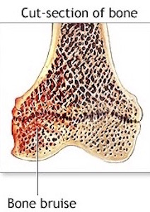
Bone bruise is a term that encompasses three different kinds of bone injuries: ⚡sub-periosteal haematoma (bleed under the outer surface of a bone) ⚡inter-osseous bruising (bruising within the main structure of the bone) ⚡sub-chondral lesion (bruising under the cartilage covering a joint surface)
A bone bruise can be described as a stage before the fracture.
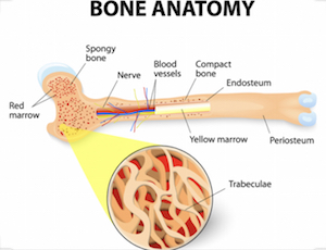
A full bone fracture occurs when all of the bone trabeculae of that specific place are fractured (broken). In the case of a bone bruise only a few of the trabeculae are broken.
EPIDEMIOLOGY/AETIOLOGY
Bone bruise injuries have been reported frequently in the knee and also in the wrist, the calcaneus, the foot, ankle and in the hip.
There are all kinds of situations where a bone bruise can occur.
The most common causes are acute injuries in the knee or ankle.
In 80% of patients with an ACL rupture a bone bruise is detected, mostly in the femur condyles or in the tibia plateau.
In the ankle bone, bruises can appear after a supination injury (sprained ankle), those will be situated in the post-lateral talus area or in the caudal tibia epiphysis (ankle joint).
A subperiosteal bleeding can also be linked to haemophilia (a disorder affecting the bloods ability to clot). This can occur together with osteolysis.
CHARACTERISTICS/CLINICAL PRESENTATION
There are 3 different kinds of bone bruise:
- Sub-periosteal haematoma
This is a concentrated collection of blood underneath the periosteum of the bone. It will appear mostly after a direct high-force trauma on the bone. This type is most common in the lower extremities. - Inter-osseous bruising
This is a damage of the bone marrow. The blood supply within the bone is damaged, and this causes internal bleedings. The trigger to this type of bone bruise is a repetitive high compressive force on the bone (extreme pressure on regular base).
The areas most affected are the knee and the ankle from professional athletes, such as footballers, basketball players and runners. - Sub-chondral lesion
This type will occur beneath the cartilage layer of a joint. The main trigger is an extreme compressive force that literally crushes the cells, that results in a separation of the cartilage (or ligament) and the underlying bone, plus bleeding when the energy of the impact extends into the bone. The other trigger is a shearing force, it sustains from a rotational mechanism such as twisting and translational forces. These will also cause that the cartilage tissue will be stripped away and exposing the underlying bone. It results in the same injury as a compressive force injury but this is another source of the injury. This type is seen more frequently in foot- and basketball players.
For the three types: an extreme compressive force can include jumping or the impact from running on hard surfaces.

DIAGNOSTIC PROCEDURES
Bone bruises do not show up on X-rays, but an X-ray can confirm that a fracture is not present.
The diagnosis of a bone bruise is mainly based on T2-weighted fat-suppressed images or T1-weighted imaging (MRI Scans).This is the best way to find out whether the patient suffers from a bone bruise.
EXAMINATION
The history of the injury will often lead your physio to suspect a bone bruise has occurred. It is often accompanied by other injuries including ligamentous sprains and cartilage injuries.
There are a lot of studies that investigate the connection between an ACL(Anterior Cruciate Ligament in the knee) tear and a bone bruise of the knee. They identified bone bruising as the most important secondary signs for the diagnosis of ACL injury. In the same studies they have also investigated the connection between MCL(Medial Collateral Ligament in the knee) tear, medial meniscus (cartilage) tear and bone bruising.:(Vellet AD et al., Mink JH. et al 1993, Mink et al 1989, Rosen MA et al)
Another study was investigating bone bruise appearance after supination injury of the ankle. This study showed that there was no significant relation between an ligament injury and a bone bruise. The bone bruises in the ankle are common in uncomplicated injuries and have minor, if any, clinical significance.
Patients with a bone bruise seem to have protracted clinical recovery, with more effusions and pain at matched time intervals and a slower return of motion.
CAN PHYSIO HELP?
- If your physio suspects you have a bone bruise they will be able arrange the necessary scans to investigate further.
- The treatment of a bone bruise consists of rest and avoiding overload of the injured bony areas.
- Repetitive or heavy loading of the affected area must be avoided and crutches used when necessary to avoid aggravation of the problem.
- Usually only painkillers (such as a small dosage of ibuprofen) are given to relieve the pain.
- The patient should receive advice about how he could reduce the load on the affected area and be made clear that if he does not rest enough the healing process will slow down or the structure can be damaged even more.
- Non-weight bearing and partial weight bearing exercises can be given to maintain strength in the affected area & improve range of movement.
- Treatment for any associated ligamentous or meniscal injuries.
- Advice regarding a gradual return to activity
- A rehabilitation programme to return to sport should be provided, monitored and progressed.
- Contact us now to arrange a consultation.
- The time for the resolution of a bone bruise is variable. At its earliest the bruise will be gone 3 weeks after the acute trauma. In all cases the bone bruises disappeared at 2 years after the trauma.
REFERENCES
- V. Mandalia, A.J.B. Fogg, R. Chari, J. Murray, A. Beale, J.H.L. Henson. Bone bruising of the knee. Clinical Radiology 2005; 60, 627–636 Grades of recommendation:A
- V. Mandalia, J.H.L. Henson. Traumatic bone bruising – A review article, European Journal of Radiology 2008; 67; 54–61 Grades of recommendation A
- Janice Polandit, 5 Things You Need to Know About a Bone Bruise, 2011; http://www.livestrong.com/article/5521-need-bone-bruise/ Grades of recommendation F
- L.C. Jungueira and J. Carneiro, “Functional Histology” 2010; 167
- Christoph Rangger, Anton Kathrein, Martin C Freund, et al. Bone Bruise of the Knee. Acta Orthop Scand 1998; 69(3) : 291-294. Grades of recommendation B
- Ville Alanen, Simo Taimela, Jaakko Kinnunen, Seppo K. Koskinen, Erkki Karaharju. Incidence and clinical significance of bone bruises after supination injury of the ankle. J Bone Joint Surg [Br] 1998;80-B:513-5. Grades of recommendation B
- Prof. Dr. S. Van Creveld and Dr. M. Kingma Subperiostal haemorrhage in haemophilia A and B. Ned. T. Geneesk. 105. I. 22. 1961; 1095-1098 Grades of recommendation F
- Simone S. Boks, Dammis Vroegindeweij, Bart W. Koes, et al. MRI Follow-Up of posttraumatic Bone Bruises of the knee in General Practice. AJR 2007; 189:556–562 Grades of recommendation B


Nancy Dominguez
Is it possiblr to have a none bruise following a knee replacement? A dark bruise occurred during post op at the outside of the knee below the kneecap about two-3inches in length and maintains with swelling and a great amt. of pain. I’m a year and a half post op , the knee grinds and pops when walking. I also need the hip replaced on the same leg. I know there can b referred pain, but the dark almost black bruise has stayed consistent. Since I can’t do an mri with an implant, will a bone scan work to show if there is a bruised bone ? Possible followup protocol?
Bradley Physio
Hi Nancy – Thank you for your enquiry. This discolouration doesn’t sound like typical bone bruising but is possibly due to circulatory changes flowing your surgery. I would recommend that you show your doctor and ask his opinion.
Rebecca Spear
Hi, I had an MRI done for my knee injury a year ago, thinking it would be a meniscus tear but it turned out to be a bruised bone. I have now had this injury for four years and it shows no sign of healing – if anything it’s getting worse and affecting other areas of my life such as work and exercising. After I got the results from the MRI, the Doctor basically said I’d have to deal with the chronic pain and gave no further advice. Do you have any advice as to what I should do and how to move forward with medical professionals?
Bradley Physio
Hi Rebecca,
It sounds like we could do to have a detailed examination of your knee to decide exactly how we can help and to make sure we give you the most appropriate treatment, exercises and advice. Please give the clinic a call to arrange an appointment,
Kind regards
Tracy
Mark Hamlin
Great read!! Thanks for sharing such a great blog.
Bradley Physio
Thanks Mark!